


 Foundational characteristics of cancer include proliferation, angiogenesis, migration, evasion of apoptosis, and cellular immortality. Find key markers for these cellular processes and antibodies to detect them.
 The SUMOplot™ Analysis Program predicts and scores sumoylation sites in your protein. SUMOylation is a post-translational modification involved in various cellular processes, such as nuclear-cytosolic transport, transcriptional regulation, apoptosis, protein stability, response to stress, and progression through the cell cycle.
 The Autophagy Receptor Motif Plotter predicts and scores autophagy receptor binding sites in your protein. Identifying proteins connected to this pathway is critical to understanding the role of autophagy in physiological as well as pathological processes such as development, differentiation, neurodegenerative diseases, stress, infection, and cancer.
DIO2 Antibody (Center)
Affinity Purified Rabbit Polyclonal Antibody (Pab)

- SPECIFICATION
- CITATIONS
- PROTOCOLS
- BACKGROUND

Application
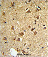
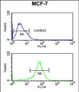
| WB, FC, IHC-P, E |
|---|---|
| Primary Accession | Q92813 |
| Other Accession | Q6QN12 |
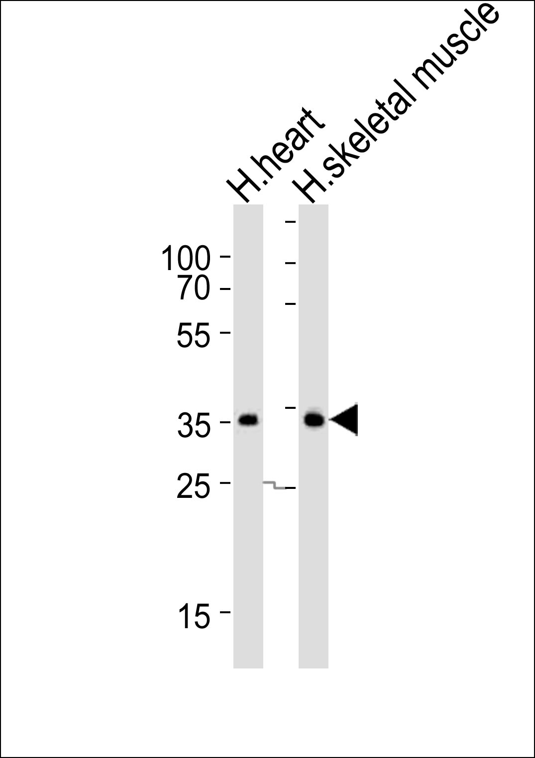
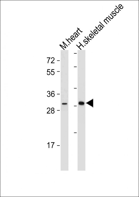
| Reactivity | Human, Mouse |
| Predicted | Pig |
| Host | Rabbit |
| Clonality | Polyclonal |
| Isotype | Rabbit IgG |
| Calculated MW | 30552 Da |
| Antigen Region | 165-191 aa |
| Gene ID | 1734 |
|---|---|
| Other Names | Type II iodothyronine deiodinase, 5DII, DIOII, Type 2 DI, Type-II 5'-deiodinase, DIO2, ITDI2, TXDI2 |
| Target/Specificity | This DIO2 antibody is generated from rabbits immunized with a KLH conjugated synthetic peptide between 165-191 amino acids of human DIO2. |
| Dilution | WB~~1:500-2000 FC~~1:10~50 IHC-P~~1:50~100 E~~Use at an assay dependent concentration. |
| Format | Purified polyclonal antibody supplied in PBS with 0.09% (W/V) sodium azide. This antibody is purified through a protein A column, followed by peptide affinity purification. |
| Storage | Maintain refrigerated at 2-8°C for up to 2 weeks. For long term storage store at -20°C in small aliquots to prevent freeze-thaw cycles. |
| Precautions | DIO2 Antibody (Center) is for research use only and not for use in diagnostic or therapeutic procedures. |
| Name | DIO2 |
|---|---|
| Synonyms | ITDI2, TXDI2 |
| Function | Plays a crucial role in the metabolism of thyroid hormones (TH) and has specific roles in TH activation and inactivation by deiodination (PubMed:12586771, PubMed:11108274, PubMed:10403186, PubMed:18821722). Catalyzes the deiodination of L-thyroxine (T4) to 3,5,3'-triiodothyronine (T3), 3,3',5'-triiodothyronine (rT3) to 3,3'- diiodothyronine (3,3'-T2) and 3',5'-diiodothyronine (3',5'-T2) to 3'- monoiodothyronine (3'-T1) via outer-ring deiodination (ORD) (PubMed:12586771, PubMed:11108274, PubMed:10403186, PubMed:18821722, PubMed:18339710). Catalyzes the phenolic ring deiodinations of 3,3',5'- triiodothyronamine and 3',5'- diiodothyronamine (PubMed:18339710). |
| Cellular Location | Endoplasmic reticulum membrane; Single-pass type III membrane protein |
| Tissue Location | Isoform 1 is expressed in the lung, trachea, kidney, heart, skeletal muscle, placenta, fetal brain and several regions of the adult brain (PubMed:11165050, PubMed:8755651). Isoform 2 is expressed in the brain, heart, kidney and trachea (PubMed:11165050) |

Thousands of laboratories across the world have published research that depended on the performance of antibodies from Abcepta to advance their research. Check out links to articles that cite our products in major peer-reviewed journals, organized by research category.
info@abcepta.com, and receive a free "I Love Antibodies" mug.

Provided below are standard protocols that you may find useful for product applications.
Background
DIO2 belongs to the iodothyronine deiodinase family. It activates thyroid hormone by converting the prohormone thyroxine (T4) by outer ring deiodination (ORD) to bioactive 3,3',5-triiodothyronine (T3).
References
He,B.,et.al., Prog. Neuropsychopharmacol. Biol. Psychiatry 33 (6), 986-990 (2009)
Heemstra,K.A., et.al., J. Clin. Endocrinol. Metab. 94 (6), 2144-2150 (2009)
If you have used an Abcepta product and would like to share how it has performed, please click on the "Submit Review" button and provide the requested information. Our staff will examine and post your review and contact you if needed.
If you have any additional inquiries please email technical services at tech@abcepta.com.
Ordering Information
Other Products
Shipping Information
